Streptococcus suis outbreaks on pig farms? Mostly because pig farmers overlooked these small details
When Streptococcus suis appears on a pig farm, it is more often because pig farmers did not pay attention to these small details
Streptococcus suis disease is a zoonosis caused by infection with various pathogenic streptococci, including swine lymph node abscess and swine septicemic streptococcosis. Septicemia, purulent lymphadenitis, meningitis and arthritis are the main features of the disease. Streptococcus suis type 2 can cause meningitis, septicemia and endocarditis in humans, and in severe cases can lead to death. Streptococcus suis disease occurs in all countries with a developed pig industry. With the development of large-scale pig farming in China, Streptococcus suis disease has become a common and frequently occurring disease in pig production.
II. Epidemiology
There are many species of streptococci, which are opportunistic pathogens and are widely distributed in nature and in pig herds,
and often exist in healthy mammals and humans. Pigs are susceptible regardless of age, breed or sex, but most outbreaks occur in piglets from 3 weeks to 12 weeks of age, and peaks of disease are especially likely at weaning and when groups are mixed.
Its transmission is mainly through the mouth or respiratory tract, and it can also be transmitted vertically (some newborn piglets can be infected during farrowing). Streptococcus suis colonizes the upper respiratory tract (especially the nasal cavity and tonsils), reproductive tract and digestive tract of pigs, and the tonsil carriage rate in pigs from 4 weeks to 6 months of age is 32% to 50%.
Sick pigs and dead pigs are the main sources of infection, and subclinically healthy carrier pigs can shed the pathogen and become a source of infection, playing an important role in the infection of young pigs. The rate of pigs carrying this pathogen ranges from 0% to 100%, however, there is no obvious correlation between the pathogen carriage rate and the incidence of disease.
The prevalence of Streptococcus suis disease has no obvious seasonality and can occur throughout the year, but large-scale outbreaks are likely from July to October. Streptococcus suis infection in humans also has no obvious seasonality, and outbreaks of the disease are regional. Introducing carrier pigs from other places, mixing groups, vaccination, high temperature and high humidity, climate change, poor pen hygiene and other stress factors that reduce the animals' resistance can all induce Streptococcus suis disease. Insect vectors play an important role in the spread of the disease, transmitting the pathogen by flying between pig farms. Among the many serotypes of Streptococcus suis, type 2 is the most important pathogen in pigs and has the strongest pathogenicity. Of all Streptococcus suis isolated from the tonsils of apparently healthy pigs, as many as 50.6% are type 2, and it is also the most pathogenic to humans.
III. Clinical symptoms
Clinically, Streptococcus suis disease mainly manifests as septicemia, meningitis, arthritis and lymph node abscess. It is divided into the following types:
1. Septicemic type
It is mainly seen in the most acute cases at the beginning of an outbreak, with sudden onset and a short course, and pigs often die suddenly without any abnormal symptoms. Or there is a sudden reduction or loss of feed intake, listlessness, body temperature rising to 41 to 42°C, difficulty breathing, constipation, cyanotic conjunctiva, inability to stand, and discharge of pale red frothy fluid from the mouth and nose, with death mostly within 6 to 24 h.
Acutely affected pigs show depression, body temperature rising to 43°C, continuous fever, poor appetite, reddened conjunctiva, lacrimation, serous nasal discharge, rapid breathing, occasional coughing, and purplish red skin with hemorrhagic spots on the neck, ear margins, under the abdomen and the lower parts of the limbs, along with lameness; the course is somewhat longer, with death mostly within 3 to 5 days. The incidence rate is generally about 30%, and the mortality rate can reach 80%.
2. Meningitis type
It mostly occurs in suckling piglets and weaned piglets. At the beginning of the disease, body temperature rises to 40.5 to 42.5°C, with cessation of feeding, constipation and serous and mucous nasal discharge, and neurological symptoms appear, manifested as incoordination, blind walking, circling, empty chewing, teeth grinding, lying on the back, paralysis of the hindquarters, lying on the side on the ground and paddling of the limbs, resembling swimming. The acute type mostly dies within 30 to 36 h.
The subacute or chronic type has a somewhat longer course and mainly manifests as polyarthritis, with gradual emaciation and death from exhaustion, or recovery.
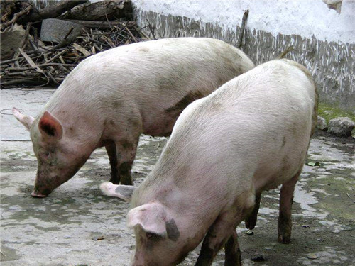
3. Arthritis type
It is mainly converted from the first two types, or manifests as arthritis from the onset of disease. Affected pigs have swelling, pain and lameness in one or several limb joints and cannot stand, with a course of 2 to 3 weeks.
4. Lymph node abscess type
This type is caused by Streptococcus suis infection through the mouth, nose and skin wounds. It is more common in weaned piglets and finishing pigs sent to market, spreads slowly and has a low incidence rate, but once it occurs in a herd, it is very difficult to eliminate.
It mainly manifests as suppuration and abscess formation in the lymph nodes under the jaw, in the pharynx and in the neck. The affected lymph nodes first develop small abscesses, which then gradually enlarge; 3 weeks after infection there is marked local swelling that feels hard on palpation and is hot and painful. Affected pigs have difficulty with feeding, chewing, swallowing and breathing. After the abscess matures, the epidermis becomes necrotic and ruptures, discharging pus. After the pus is drained completely, the systemic symptoms are significantly reduced, and granulation tissue grows and heals with scarring. The course is 3 to 5 weeks.
IV. Pathological changes
1. Acute septicemic type
Poor blood clotting, purple patches on the skin, and hemorrhage in the mucous membranes, serous membranes and subcutis. The nasal mucosa is purplish red, with congestion and hemorrhage. The larynx and trachea are congested, and large amounts of foam are commonly seen. The lungs are congested and swollen. The systemic lymphatic vessels show varying degrees of enlargement, congestion and hemorrhage. There is pericardial effusion, pale yellow, and a few cases show mild fibrinous pericarditis, with hemorrhagic spots on the endocardium. Some cases show mild fibrinous pleurisy. There is a small amount of pale yellow effusion in the abdominal cavity, and some cases have relatively mild fibrinous peritonitis. In most cases the spleen is enlarged, in a few cases enlarged 1—3 times, dark red or purplish blue, soft and easily torn. The mucosa of the stomach and small intestine shows varying degrees of congestion and hemorrhage. The kidneys are mostly mildly enlarged, congested and hemorrhagic. The meninges show varying degrees of congestion, sometimes with hemorrhage.
2. Meningitis type
The meninges are congested and hemorrhagic, with hemorrhage in severe cases, and in a few cases the subarachnoid space is filled with effusion; on the cut surface of the brain, obvious small petechial hemorrhages can be seen in the white matter and gray matter; other changes are similar to those of the septicemic type.
3. Chronic type
In endocarditis, the heart valves are thickened with a rough surface, and cauliflower-like vegetations are present on the valves, commonly on the bicuspid or tricuspid valve, and sometimes also in the atria, ventricles and blood vessels. In arthritis, there is yellow jelly-like fluid or fibrinous purulent material inside and outside the joint capsule.
V. Diagnosis and differentiation
Based on the epidemiological characteristics, symptoms and pathological changes of the disease, only a preliminary diagnosis can be made; confirmation requires laboratory diagnosis.
1. Microscopic examination of smears is a relatively simple diagnostic method. Samples such as liver, spleen, lymph nodes, blood, joint fluid and pus from affected pigs can be taken, smeared, stained and examined microscopically to observe whether typical streptococci are present.
2. Isolation and culture of bacteria. Aseptically collect samples such as liver, spleen, lymph nodes, blood, joint fluid and pus and inoculate them onto blood agar plates, and observe colony growth, whether hemolysis occurs, and the type of hemolysis. If β-hemolysis appears in the colonies, further bacterial morphological and biochemical identification is carried out. Serological and typing identification can also be further performed. PCR is used to identify the virulence factors of the strain.
3. Animal inoculation test. Inoculate the collected samples or bacterial cultures into rabbits, mice and pigeons, observe the incidence of disease in the test animals, and recover and identify the bacteria from dead animals.
In differential diagnosis, this disease should mainly be distinguished from listeriosis, swine erysipelas, swine paratyphoid and swine fever.
VI. Comprehensiveprevention and control
1.treatmentinformation
Isolate the affected pigs and carry out correspondingtreatment according to the different disease types. For lymph node abscess, once the abscess has matured (softened), incise it promptly, drain the pus, rinse with 30% hydrogen peroxide or 0.1% potassium permanganate, and then apply iodine tincture.
For the septicemic type or meningitis type, antibiotics or sulfonamides should be used early and in large doses. Penicillin 400,000—1,000,000 units per pig per time, intramuscularly 2—4 times daily; gentamicin 1—2 mg per kg body weight, intramuscularly twice daily. Acetylciprofloxacin can also be used fortreatment, 2.5—10 mg per kg body weight, injected once every 12 hours for 3 consecutive days, which can rapidly improve symptoms, with efficacy clearly superior to penicillin.
2. Prevention information
(1) Isolate affected pigs and eliminate the source of infection. Carcasses of suspected affected pigs found after slaughter may be eaten only after high-temperature treatment. Remove sharp objects from the pig pens.
(2) Immunoprevention In epidemic areas (farms), the first immunization is given at 60 days of age by vaccinatingbreeding pigswith Streptococcus suis aluminum hydroxide gel vaccine, and thereafter immunized once each in spring and autumn every year; regardless of size, all pigs are injected intramuscularly or subcutaneously with 5 ml, or 3 ml of concentrated vaccine; immunity develops 21 days after injection and the immune period is about 6 months. For attenuated Streptococcus suis vaccine, each pig is injected intramuscularly or subcutaneously with 1 ml, immunity develops in 14 days, and the immune period is six months.
(3) Drug prevention After this disease occurs on a pig farm, if the vaccine cannot be purchased temporarily, drugs can be used for prevention toprevent and controlthe occurrence of this disease. Add 125 g of tetracycline per ton of feed and feed continuously for 4—6 weeks.
3. Public health
(1) Strengthen quarantine of live pigs. Any affected pigs detected before slaughter should be urgently isolated fortreatment, and may be slaughtered only two weeks after recovery; those with suspicious lesions found after slaughter should undergo harmless treatment. Emergency-slaughtered pigs should be handled in a separate emergency slaughter room to prevent contamination of healthy pork. Emergency-slaughtered affected pigs may not be sold without harmless treatment. The swill from the slaughterhouse must be boiled before being fed. At the same time, strengthen the disinfection and hygiene systems of slaughterhouses and live pig trading markets.
(2) People should stay away from diseased animals, not eat pork from dead or diseased pigs or diseased pork that has not undergone harmless treatment, not directly contact dead or diseased animals, and pay attention to disinfection in operations such as castration, injection and delivery with umbilical cord cutting.
(3) Once the disease breaks out and spreads, according to the Animal Epidemic Prevention Law, emergency isolation, blockade and measures should be immediately taken to promptly control and stamp out the animal epidemic.