Detection Methods and Case Analysis of Porcine Circovirus Disease
Porcine circovirus disease refers to a general term for a series of diseases with porcine circovirus type 2 (PCV) as the main pathogen, either alone or as a secondary or mixed infection with other pathogenic microorganisms. It is the smallest animal virus discovered to date.
Porcine circovirus disease, also known as postweaning multisystemic wasting syndrome (PMWS), is a type of viral infectious disease characterized mainly by physical decline, emaciation, diarrhea, respiratory difficulty, immunosuppression, and secondary infection by multiple pathogens.
I. Pathogen
Porcine circovirus (PCV) is a member of the genus Circovirus in the family Circoviridae. This virus is the smallest among animal viruses, with a particle diameter of 14 to 25 nanometers, icosahedral symmetry, no envelope, and a genome consisting of single-stranded circular DNA.
Three serotypes of PCV are currently known, namely PCV1, PCV2, and PCV3. PCV1 is a non-pathogenic virus, while PCV2 is a pathogenic virus.
II. Epidemiological Characteristics
This disease can occur in all four seasons. Postweaning multisystemic wasting syndrome (PMWS) mainly affects piglets 2 to 3 weeks after weaning, but in pig farms practicing early segregated weaning, weaned pigs at 10 to 14 days of age also develop the disease. The virus can be shed in feces and nasal secretions, transmitted through the digestive tract, and may also be transmitted vertically to piglets through the placenta. The morbidity and mortality rates vary. During endemic outbreaks, both morbidity and mortality rates are relatively low, but during acute outbreaks, the morbidity rate can reach 50%, and the mortality rate can reach 20% to 30%.
III. Clinical Symptoms
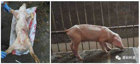
It mainly manifests in piglets at 6 to 8 weeks of age. Around weaning, especially beginning two weeks after weaning, piglets develop skin lesions, namely large numbers of red and purplish-red spots appearing on the back, at the base of the ears, and even on the abdomen. These spots generally protrude outward and are relatively hard in texture. The proportion of runt pigs in nursery pens increases significantly, manifesting as poor appetite, weakness, weight loss, sometimes coughing or central nervous system disorders, and even respiratory difficulty, jaundice, and enlarged lymph nodes (which can be palpated).
IV. Necropsy Symptoms
Necropsy reveals generalized lymph node enlargement, especially enlargement of the inguinal lymph nodes, mediastinal lymph nodes, hilar lymph nodes, mesenteric lymph nodes, and submandibular lymph nodes, with increased firmness on cut surface and a uniform white appearance; some lymph nodes show hemorrhagic and suppurative lesions. The lungs are swollen, firm, or rubbery; in severe cases, there are hemorrhagic spots in the alveoli; some apical and cardiac lobes of the lungs are atrophied or show substantive lesions. The liver is dark and atrophied, with proliferation of connective tissue between hepatic lobules. The spleen is abnormally enlarged and shows meat-like changes. The kidneys are edematous and grayish-white, sometimes with white necrotic foci beneath the capsule. The mucosa of the esophageal region of the stomach shows edema and non-hemorrhagic ulcers. The intestinal wall of the ileum and colon segments becomes thinner, and the cecal and colonic mucosa show congestion and hemorrhage.
V. Clinical Diagnosis
Reliable conclusions in clinical diagnosis must be obtained by combining clinical symptoms, pathological changes, and laboratory pathogen or antibody detection. The two commonly used methods are ELISA and PCR monitoring.
1. ELISA diagnostic method: It can be used to detect the level of viral antibodies in serum. It monitors the immune level of circovirus vaccines in pig farms and helps formulate reasonable immunization programs.
2. PCR method: It is a rapid, simple, and specific diagnostic method. Using PCV-specific or group-specific primers to perform gene amplification from the tissues of diseased pigs, PCV infection is confirmed based on the restriction enzyme digestion pattern and base sequence of the amplified products.
VI. Prevention and Control Measures
1. Persistent infection with PCV2 makes this disease more economically devastating. The application of antibiotics and good management help solve the problem of concurrent infections.
2. Strengthening feeding management, reducing stocking density, implementing a strict all-in-all-out system and mixing-group system, reducing environmental stress factors, controlling concurrent infections, ensuring a stable immune status of the herd, strengthening internal and external biosecurity measures on the farm, and ensuring that purchased pigs come from clean farms are effective measures to prevent and control this disease and reduce economic losses.
3. Do a good job in farm sanitation, ventilation, and disinfection.
In May 2017, the Zhongnong Veterinary Testing Center received diseased material submitted by a farmer in Zhuozhou, Hebei. According to the description of the farm manager, the testing center conducted several laboratory tests. The specific content and results are as follows:
1. Report information and test report number
2. Test items: PCR testing for swine fever, porcine reproductive and respiratory syndrome, and circovirus, as well as bacterial testing and drug sensitivity testing on organs.
3. Test results: The PCR test result was positive for circovirus type 2 and negative for the others.
Drug sensitivity test results: as shown in the table below
4. Clinical recommendations: Based on the test results, expert Teacher Yin from the testing center formulated a reasonabletreatmentplan.