This article describes the main lesions of the principal bacterial pneumonias. Although most are opportunistic agents, two primary bacteria can cause lesions on their own.
The bacteria that cause pneumonia in pigs and can cause lesions on their own can be classified as primary pathogens. These include Mycoplasma hyopneumoniae, Actinobacillus pleuropneumoniae, Bordetella bronchiseptica and Pasteurella multocida. Secondary pathogens are bacteria that act after a previous infection by a primary pathogen or in immunocompromised animals; most of these are Pasteurella multocida, Streptococcus suis, Glaesserella (formerly known as Haemophilus parasuis) and Trueperella pyogenes.
Among the primary pathogens, Mycoplasma hyopneumoniae is the agent that causes enzootic pneumonia and can cause serious economic losses on pig farms, being associated with decreased average daily gain, increased feed conversion ratio and increased mortality due to secondary infections. This prokaryotic cell belongs to the class Mollicutes, is highly pleomorphic, and is characterized by the lack of a cell wall, circular double-stranded DNA, a polysaccharide capsule with radial fibers, and being very difficult to culture.
To exert its pathogenic effect, it binds to the cilia of the respiratory epithelial cells, causing loss of cilia and altered function of the mucociliary system, which means that pulmonary secretions, inhaled particles and pathogens entering the airway cannot be cleared, so they move down the lower respiratory tract, complicating the respiratory process. In the lung, this bacterium can cause bronchointerstitial pneumonia, which macroscopically appears as red consolidation in the cranioventral part of the lung, clearly demarcated from the other parenchymal areas (Figure 1). Under the microscope, peribronchial, peribronchiolar and perivascular lymphoid tissue hyperplasia can be observed, which is characteristic of this process (Figure 2).
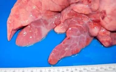
Figure 1: Bronchointerstitial pneumonia caused by Mycoplasma hyopneumoniae
Figure 2: Peribronchiolar lymphoid tissue hyperplasia caused by Mycoplasma hyopneumoniae.
Actinobacillus pleuropneumoniae (APP) is an encapsulated Gram-negative coccobacillus with two biotypes: biotype I, which depends on nicotinamide adenine dinucleotide (NAD), and biotype II, which does not depend on nicotinamide adenine dinucleotide. There are 18 serotypes in total: 1-12 and 15-18 belong to biotype I, while serotypes 13 and 14 belong to biotype II. Actinobacillus pleuropneumoniae can produce 4 toxins: ApxI, II and III, which are specific to certain strains, and ApxIV, which is common to all strains, which is why ApxIV is the target of several indirect ELISA diagnostic tests. This bacterium colonizes the tonsils and moves down to the lower respiratory system, adhering to alveolar pneumocytes, where it secretes toxins, producing hemolytic and cytotoxic effects and altering endothelial cell function; phagocytosis by alveolar macrophages will lead to activation of the coagulation cascade, formation of microthrombi and phenomena of ischemic necrosis, which are the most characteristic features of this pathogen.
The type of pulmonary lesion it causes is fibrinous bronchopneumonia. In peracute and acute cases, the lesions occur mainly in the dorsal and distal parts of the lung (Figure 3), with coagulative necrosis (infarction) and hemorrhage, friable, and marbled on cut surface. Microscopically, fibrin and degenerated leukocytes are seen in the alveolar spaces, bronchi and bronchioles, with necrosis and hemorrhage of varying size, alveolar and interstitial edema, and fibrinous pleuritis. These same lesions may be produced by some highly virulent strains of Pasteurella multocida, so to help us make a correct diagnosis, we need to analyze the clinical history, macroscopic lesion presentation and microbiology.
Actinobacillus pleuropneumoniae can also occur with chronic lesions; acute lesions cause hematogenous spread, and white multifocal lesions of varying size can be seen in the lung.
Figure 3: Caudal lobe lesions produced by Actinobacillus pleuropneumoniae
Most Pasteurella multocida strains are secondary pathogens, and they can cause lesions ranging from fibrinous bronchopneumonia to catarrhal purulent bronchopneumonia. The former produces fibrinous exudate within the alveoli, together with fibrin deposition on the pleural surface. In addition, due to thrombosis in the blood vessels, coagulative necrosis appears in the lung. In catarrhal purulent bronchopneumonia, mucous exudate or catarrhal purulent pneumonia appears in the airways (trachea, bronchi and bronchioles) and alveolar spaces (Figure 4). Another factor causing this type of bronchopneumonia, especially in the first weeks after birth, may be bronchial septicemia, which may also be accompanied by non-progressive atrophic rhinitis.
Figure 4: Purulent bronchopneumonia: enlarged cranioventral consolidation, with small yellow-white areas visible (indicated by arrows) corresponding to alveoli filled with purulent material. Interstitial edema can also be observed.
Bacteria such as Trueperella pyogenes and Streptococcus suis may be associated with embolic pneumonia, related to omphalophlebitis, bacterial infections of the skin or hooves (Trueperella pyogenes), or related to endocarditis (Streptococcus suis); in these cases, the bacteria travel from these lesions through the blood to the lungs. Macroscopically, multifocal lesions are visible distributed throughout the lung, whitish, between 1 mm and 2 cm in diameter, surrounded in the most severe cases by pale red rings, and rapidly progressing to abscesses (Figure 5).
Figure 5: Embolic pneumonia caused by Trueperella pyogenes.
Figure 6: Fibrinous pleuritis observed at the slaughterhouse.