How to diagnose and treat bovine viral diarrhea? A highly harmful infectious disease that must not be ignored
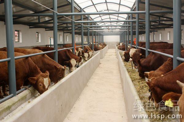
Cattle farming
Bovine viral diarrhea is an infectious disease caused by bovine viral diarrhea virus. Cattle of all ages are susceptible, with young calves being the most susceptible. It can be transmitted through direct or indirect contact with the secretions, excretions, blood and spleen of infected cattle.
1. Symptoms
The incubation period of the disease is 7~14 days.
1.1 Acute form Sudden onset, with body temperature rising to 40°C~42°C, lasting only 2~3 days, and some animals show a second rise (biphasic fever). As body temperature rises, white blood cells decrease, lasting 1~6 days, followed by a slight increase in white blood cells. Sick cattle are anorexic, have nasal discharge, salivate, and their breath has a foul odor. Severe diarrhea usually occurs after damage to the oral mucosa, initially watery diarrhea, later containing mucus and blood. Some cases often have erosion and necrosis of the hoof laminae and interdigital skin, leading to lameness.
Acute infection causes severe immunosuppression in cattle for 7~14 days or until the recovery period, during which they are highly susceptible to secondary infections. Infection with harmful bacteria (such as Pasteurella, Haemophilus somnus, Mycoplasma, etc.) can easily cause secondary conditions (such as pneumonia).
When cows are infected with this disease during pregnancy, the virus also invades the fetus through the placenta, causing abortion or congenital defects in calves, the most common being cerebellar hypoplasia (i.e., infection at 100~200 days of gestation). Affected calves show only mild ataxia or completely lose coordination and the ability to stand.
1.2 Chronic form Fewer cases show obvious symptoms, but body temperature may fluctuate above normal. The most notable symptoms are erosion on the nasal mirror, serous ocular discharge, gingivitis, laminitis, and erosion and necrosis of the interdigital skin. Generally there is no diarrhea.
2. Pathological changes
30%~50% of diseased cattle have erosion of the oral mucosa (gingiva, palate, both sides of the tongue surface and buccal mucosa), 80% of affected cattle with clinical symptoms have erosion and superficial ulcers on the nasal mirror and nostrils, and severe cases have ulcers and diffuse necrosis of the pharyngeal and laryngeal mucosa. The characteristic lesion is erosion spots of varying sizes arranged in straight lines on the esophageal mucosa. The rumen, abomasum and intestines all show varying degrees of edema, hemorrhage, ulcers and necrotic inflammation. Aborted fetuses also have digestive tract damage.
3. Diagnosis
During an outbreak of this disease, a preliminary diagnosis can be made based on its history, symptoms and pathological changes. However, fewer than 50% of diseased cattle show clinical symptoms and lesions, and most cattle show subclinical or mild infection, making diagnosis somewhat difficult. Confirmation requires virus isolation and identification and serological testing.
4. Treatment
Mild cases do not require special treatment, but fresh feed and water should be provided, and external stress, transport or vaccination should be avoided. For cattle with clinical specific symptoms such as mucosal erosion and diarrhea, symptomatic treatment can be given, that is, supplementing water and electrolytes, protecting the gastrointestinal mucosa, and using antibiotics for prevention to reduce the possibility of secondary infection by opportunistic bacteria.
5. Prevention
5.1 Identify and cull persistently infected cattle, because this type of cattle is not easy to detect, but they always carry the virus and continuously shed it through secretions.
5.2 Strictly prohibiting the purchase of untested cattle can effectively reduce the risk of introducing the virus.
5.3 Immunize with inactivated vaccine. Initial immunization requires at least 2 doses, with an interval of 30 days, to ensure sufficient immunity is produced.