What are the traditional Chinese medicine treatments for extralobar pulmonary sequestration?
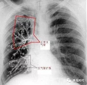
Extralobar means that pulmonary sequestration forms outside the lung lobe, and it is separated from the normal lung lobe by the visceral pleura, that is, the sequestered lung outside the lung lobe is completely wrapped by the visceral pleura. Another characteristic is that it usually occurs together with other malformations. It is commonly combined with left diaphragmatic hernia, accounting for 30%, and is also often combined with cardiovascular malformations, pectus excavatum, bronchogenic cyst, esophagobronchial fistula, etc. In terms of blood supply, extralobar sequestered lung receives arterial supply, most commonly from the abdominal aorta. Venous return is most commonly through the inferior vena cava, portal vein, or azygos vein.
In terms of treatment, extralobar pulmonary sequestration is most suitable for Western medicine surgical treatment when permitted. However, whenever there is surgery, traditional Chinese medicine says it will damage vital qi, so on the premise of not affecting physiological function and growth and development, infants and young children with extralobar pulmonary sequestration do not need surgery. Moreover, generally speaking, for both intralobar and extralobar pulmonary sequestration, in most cases symptoms appear only after infection occurs.
Clinically, extralobar pulmonary sequestration occurs in the posterior segment of the left lower lung, accounting for about 90%. The remaining about 10% often occurs under the diaphragm or within the mediastinum. Now infants are basically all born in hospitals and undergo all examinations, so it is very easy to detect. Once pulmonary sequestration is found in a newborn through imaging such as contrast studies, careful nursing must be provided, and the newborn or infant must not be allowed to contract infectious diseases such as colds.
In extralobar pulmonary sequestration, normally the sequestered lung tissue does not communicate with the adjacent bronchi, and on CT and other contrast imaging it appears as a soft tissue density mass with density normal and uniform, similar to the lung lobe; the site to pay attention to is the posterior part of the left lower lung. Extralobar sequestered lung is mostly mass-like; some are round, and some are oval or lobulated masses, which can compress surrounding organs and tissues and need to be differentiated from bronchiectasis, pneumonia, lung abscess, lung tumor, etc.