Detailed Explanation of Prevention, Control Measures and Methods for Fowl Cholera
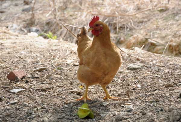
Fowl cholera is a contagious disease that affects poultry and wild birds, also known as avian pasteurellosis and avian hemorrhagic septicemia. The disease often presents with septicemic symptoms, with high morbidity and mortality, but chronic or benign courses also commonly occur.
Causative Pathogen
Pasteurella multocida is a short bacillus with bluntly rounded ends and a slightly convex center, measuring 1 to 1.5 um in length and 0.3 to 0.6 um in width. It does not form spores and is non-motile. It can be stained by ordinary dyes and is Gram-negative. When smears of diseased tissue or body fluids are stained with Wright's, Giemsa, or methylene blue and examined under the microscope, the bacteria mostly appear oval-shaped, with deep staining at both ends and lighter staining in the central portion, closely resembling two cocci arranged side by side, which is why it is also called a bipolar bacillus. In smears made from cultures, the bipolar staining is less pronounced. When stained with India ink or similar dyes, a clear capsule can be seen. Newly isolated bacteria have a wide, thick capsule, while attenuated strains that have undergone variation through artificial culture have a narrow and incomplete capsule.
Epidemiology
This disease is susceptible to various types of poultry, such as chickens, ducks, geese, and turkeys, but geese are less susceptible. Various wild birds are also susceptible. Mortality losses in chickens caused by fowl cholera usually occur in laying hen flocks, as chickens of this age are more susceptible than younger chickens. Chickens under 16 weeks of age generally have relatively strong resistance. However, outbreaks have also been observed clinically in chicken flocks at 10 d of age. The mortality rate of naturally infected chickens is typically 0 to 20% or higher, and egg production declines and persistent localized infections frequently occur. Withdrawal of feed, withdrawal of water, or sudden changes in feed can all increase the susceptibility of chickens to fowl cholera.
How fowl cholera is introduced into a chicken flock is often impossible to determine. Chronically infected birds are considered the main source of infection. Transmission of the bacteria through eggs rarely occurs. Most farm animals may be carriers of Pasteurella multocida, and contaminated cages, feed troughs, and the like can all transmit the pathogen. The spread of Pasteurella multocida within poultry flocks occurs mainly through secretions from the oral cavity, nasal cavity, and conjunctiva of diseased birds; these secretions contaminate the environment, especially feed and drinking water. Feces rarely contain live Pasteurella multocida.
Clinical Symptoms
The incubation period of natural infection is generally 2 to 9 d, and sometimes cases may suddenly break out within 48 h after the introduction of diseased chickens. Artificial infection usually causes disease within 24 to 48 h. Because the body resistance of poultry and the virulence of the bacteria vary in strength, the symptoms manifested also differ. It is generally divided into three types: peracute, acute, and chronic.
1. Peracute type: Commonly seen in the early stage of an outbreak, most frequently in high-laying chickens. Diseased chickens show no prodromal symptoms; they appear completely normal in the evening and eat their fill, then are found dead in the coop the next day.
2. Acute type: This type is the most common. Diseased chickens mainly show depression, ruffled feathers, neck retraction with eyes closed, head tucked under the wings, unwillingness to move, and standing apart from the flock in a daze. Diseased chickens often have diarrhea, passing yellow, grayish-white, or green watery droppings. Body temperature rises to 43 to 44 degrees C, with reduced appetite or anorexia and increased thirst. Breathing is difficult, and oral and nasal secretions increase. The comb and wattles turn bluish-purple, and some diseased chickens have swollen wattles with a hot, painful sensation. Laying hens stop laying. Finally, exhaustion occurs, followed by coma and death. The course of disease is as short as about half a day, or as long as 1 to 3 d.
3. Chronic type: It develops from acute cases that do not die, and is mostly seen in the late stage of an outbreak. Chronic pneumonia, chronic respiratory inflammation, and chronic gastroenteritis are relatively common. Diseased chickens have viscous secretions flowing from the nostrils, swollen sinuses, and accumulated secretions in the larynx that affect breathing. Diarrhea is frequent. Diseased chickens become emaciated, listless, and have pale combs. In some diseased chickens, one or both wattles are significantly swollen, and afterward there may be purulent caseous material, or drying, necrosis, and sloughing. Some diseased chickens have arthritis, often confined to the joints and tendon sheaths of the legs or wings, manifesting as swollen and painful joints and paralyzed toes, resulting in lameness. The course of disease may drag on for more than one month, but growth and egg production cannot recover for a long time.
Pathological Changes
Diseased chickens that die of the peracute type show no special lesions, and sometimes only a few hemorrhagic spots on the epicardium can be seen.
The lesions in acute cases are relatively characteristic. Petechial hemorrhages are commonly seen in the peritoneum, subcutaneous tissue, and abdominal fat of diseased chickens. The pericardium becomes thickened, and a large amount of opaque pale yellow fluid accumulates within the pericardial sac, sometimes containing fibrinous flocculent fluid, with hemorrhage on the epicardium and coronary fat being particularly obvious. The lungs have congestion or hemorrhagic spots. The lesions in the liver are characteristic: the liver is slightly swollen, brittle in texture, and brown or yellow-brown in color. Numerous grayish-white, pinhead-sized necrotic spots are scattered on the liver surface. The spleen generally shows no obvious changes, or is slightly enlarged with a relatively soft texture. Hemorrhage in the gizzard is significant, and the intestines, especially the duodenum, show catarrhal and hemorrhagic enteritis, with the intestinal contents containing blood.
The chronic type varies depending on the organs affected. When respiratory symptoms predominate, a large amount of viscous secretion is seen in the nasal cavity and sinuses, and in some cases pulmonary induration is observed. In cases confined to arthritis and tenosynovitis, the main findings are swollen and deformed joints, with inflammatory exudate and caseous necrosis. The wattles of roosters are swollen and contain caseous exudate. The ovaries of hens show obvious hemorrhage, and sometimes the follicles are deformed, resembling half-cooked eggs.
The pathological changes in ducks are basically similar to those in chickens. Ducks that die of fowl cholera have the pericardial sac filled with transparent orange-yellow exudate, and hemorrhagic spots on the pericardium and coronary fat. The lungs show multiple pneumonia, with occasional emphysema and hemorrhage. The nasal mucosa is congested or hemorrhagic. The liver is slightly enlarged, showing pinpoint hemorrhages and grayish-white necrotic spots. In the intestines, congestion and hemorrhage of the mucosa are most severe in the anterior segment of the small intestine and the large intestine; the posterior segment of the small intestine and the cecum are less affected. In ducklings, it manifests as multiple arthritis, mainly showing rough articular surfaces with yellow caseous material or red granulation tissue attached. The joint capsule is thickened and contains red serous fluid or grayish-yellow, turbid, viscous liquid. The liver undergoes fatty degeneration and focal necrosis.
Diagnostic Methods
A preliminary diagnosis can be made based on the epidemiology, necropsy characteristics, and clinical symptoms of diseased chickens, but a definitive diagnosis requires laboratory diagnosis. Blood smears from diseased chickens and impression smears of the liver and spleen are stained with methylene blue, Wright's, or Giemsa stain; if large numbers of short, small bacilli with bipolar deep staining are seen, this aids diagnosis. Further diagnosis requires isolation and culture of the bacteria and biochemical reactions.
Prevention and Control Measures
Strengthening the feeding and management of chicken flocks, strictly implementing veterinary hygiene and epidemic prevention measures on the farm at ordinary times, and adopting an all-in-all-out rearing system by house unit make it entirely possible to prevent the occurrence of this disease. Generally, chicken farms that have never had this disease do not carry out vaccination. Once a flock is affected, treatment measures should be taken immediately, and where conditions permit, effective drugs should be selected through drug sensitivity testing and administered to the whole flock. Sulfonamides, chloramphenicol, erythromycin, gentamicin, ciprofloxacin, enrofloxacin, and olaquindox all have relatively good efficacy. During treatment, the dosage should be sufficient and the course of treatment reasonable; after the mortality of chickens has clearly decreased, medication should be continued for another 2 to 3 d to consolidate the efficacy and prevent relapse.
In areas or chicken farms where the disease frequently occurs, the effect of drug treatment gradually decreases, and the disease is difficult to control effectively, so vaccination may be considered for prevention; however, because the immune period of the vaccine is short, the preventive and control effect is not entirely satisfactory. Where conditions permit, bacteria can be isolated from the farm itself, and after identification and qualification, an autogenous inactivated vaccine can be prepared and injected into the flock regularly. Practice has proven that through 1 to 2 years of immunization, this disease can be effectively controlled. At present, there is a relatively good fowl cholera propolis inactivated vaccine available domestically, which is safe and reliable, can be stored at 0 degrees C for two years, is easy to inject, does not affect egg production, and has no toxic side effects, and can effectively prevent and control the disease.