How to distinguish between lung bullae and pulmonary lymphangioleiomyomatosis
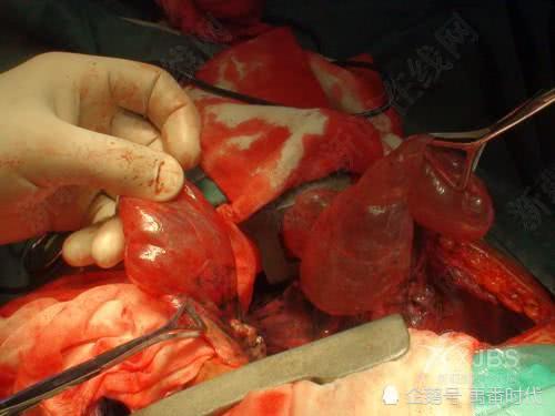
Pulmonary lymphangioleiomyomatosis, which is extremely similar to lung bullae, in its later stages, as muscle cells continuously accumulate, collagen is also deposited and increased, forming nodules, edema, or hemorrhage, and the destroyed alveoli fuse with each other to form cysts, whose morphology is very similar to lung bullae, and at this time macrophages decrease. If regular physical examinations with chest X-rays are not performed, and respiratory symptoms appear or symptoms become severe, and the patient goes to the hospital for examination, it is sometimes easily confused with lung bullae.
Lung bullae surgery
In the later stages of pulmonary lymphangioleiomyomatosis, which has already become cystic, the cyst walls are lined with flat epithelium and ciliated epithelium, and the nodules proliferating on the cysts may also extend into the cyst interior, alveoli, and bronchioles, causing the cyst walls to continuously thicken and the structure to become disordered. When dilated lymphatic vessels form lung bullae-like cysts in the muscular layer and parietal pleura that are widely distributed on the lung surface, new atypical smooth muscle cells also appear on the lymphatic vessels, bronchovascular bundles, and small veins.
These slightly shorter and pleomorphic atypical smooth muscle cells are very similar to smooth muscle and are also called clonal smooth muscle in medicine, and a certain degree of hemosiderin is present within the muscle cell tissue. During continuous growth, they also extend deep into the alveoli and inside and outside the pulmonary interstitium, causing luminal obstruction and dilation, and forming new diffuse terminal airspace dilated cysts with a diameter of 1 cm or larger. During continuous development, the pulmonary lymphatic vessels become dilated or twisted, and rupture of small veins causes a small amount of bleeding; patients may cough up blood, and may even develop hemopneumothorax.
Pulmonary lymphangioleiomyoma surgery
Pulmonary lymphangioleiomyomatosis also easily leads to involvement and enlargement of the lymph nodes in the hilum, mediastinum, and posterior abdominal wall, and may also cause thickening and dilation of the thoracic duct, with internal obstruction forming chylothorax, and even more severely, extrapulmonary organ or tissue vascular leiomyomatosis. For example, vascular leiomyomatosis occurs intermittently or simultaneously in the kidneys, posterior abdominal wall, abdominal cavity, pelvic cavity, and other sites, and the body shows corresponding organ- or tissue-related symptoms. In addition, the associated symptoms of bullae and pulmonary lymphangioleiomyomatosis are also different, and combining them makes differentiation easier.
If you have questions or need help, you can call 01062170649 13269852331