Fish gills growing mold: is there still a chance to save them? Proper treatment is key
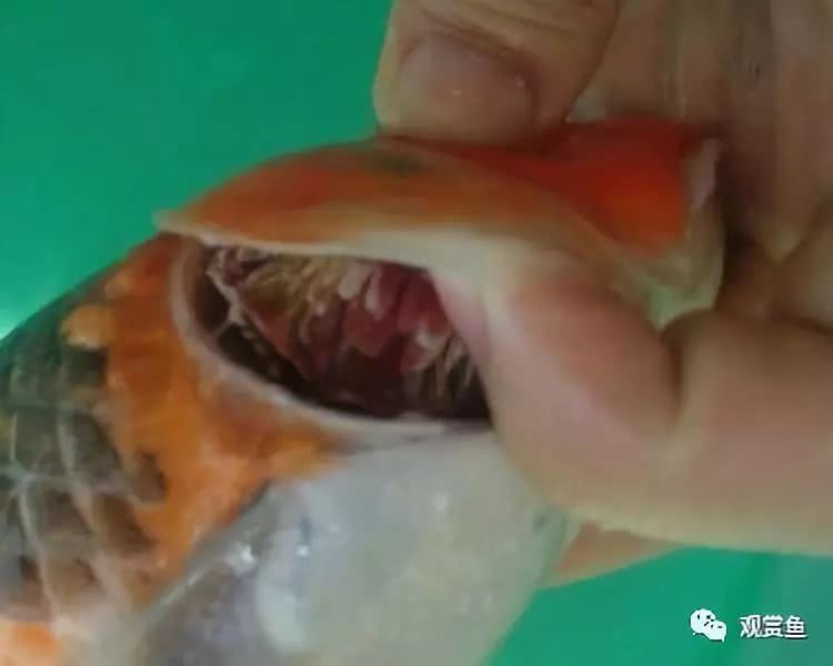
On May 19, the editor published an article titled "Fish gills turning black, red, and rotting away: these all need attention, or they may accidentally infect other fish," discussing the problem of gill rot in ornamental fish. I believe many fish keepers have encountered fish gill rot disease or water mold disease, but the one we are talking about today is a bit different from the previous two. Water mold disease outbreaks are systemic, and the early stage of gill rot is similar to gill mold. The two do not need to be distinguished too much, and the same treatment can be used for both. Today we will talk about gill mold disease.
Gill mold disease is caused by gill mold parasitizing the gills of ornamental fish. It mainly harms koi and goldfish from the fry stage to the fingerling stage, and mostly occurs when water quality deteriorates, especially in water bodies with very high organic matter content. Once a fish becomes ill, it is mostly acute, and it can die within 1-2 days. The disease is prevalent in the summer and autumn seasons from May to October each year, especially from May to July. In other seasons, as the water temperature decreases, it turns into subacute and chronic forms.
The pathogen of this disease is gill mold, mainly Branchiomyces sanguinis and Branchiomyces demigrans, which can produce non-septate branched hyphae and endospore hyphae. The hyphal body diameter is 6.6-21.6 micrometers, and the spores are smaller in diameter, 4.8-8.4 micrometers, with an average of 6.6 micrometers. The spores can live in water without a host and can directly invade the fish body or be ingested by fish, then enter the blood circulation through the intestinal lymphatic vessels, and finally germinate in the gill blood vessels and develop into hyphae. The hyphae continuously extend inward and branch again, penetrating the tissues, growing along the branches of the gill filament blood vessels or piercing into the cartilage, destroying the gill tissue, causing blood circulation disorders, and forming blood clots.
Main symptoms
The gill filaments of the diseased fish lose their normal bright red color and become pink or pale. When viewed against the light, punctate or patchy congestion can be seen on the gill filaments, resembling marble and presenting a mottled gill appearance. The gill filaments are obviously thickened, and the secondary lamellae are covered by proliferated and enlarged epithelial cells. As the disease progresses, the injured tissue rots. The diseased fish has reduced appetite, swims slowly, has increased mucus on the gills, and experiences breathing difficulty. It disperses and floats on the water surface, then swims wildly in 2-3 circles, after which the fish body loses balance with the head pointing downward. This repeats several times, and death occurs due to breathing difficulty.
According to the severity of the disease, gill mold disease can be divided into three types: acute, subacute, and chronic.
Acute gill mold disease is mostly acute. After onset, mass death begins in 3-5 days and lasts 5-10 days, with a mortality rate above 60%.
The subacute course is longer, the necrotic parts can still regenerate, and the mortality rate is 10%-40%.
Chronic symptoms are obvious. Sometimes a small part of the gills is necrotic, and in individual cases it appears pale. In some diseased fish, the ends of the gill filaments show swelling.
How to judge?
On-site diagnosis: A preliminary diagnosis can be made based on clinical symptoms and pathological changes, but further laboratory diagnosis is needed for confirmation.
Laboratory diagnosis: Cut a small amount of rotten gill filaments, add a coverslip, and observe under a microscope to see whether branched hyphae are present, and a diagnosis can be made. If conditions permit, analyze the water quality of the fish pond to see whether there is a large amount of organic matter and water quality deterioration.
Key prevention points
Always maintain excellent and stable water quality, add new water in a timely manner, and avoid excessive organic matter in the water, which can reduce the chance of disease.
Strengthen feeding management and feed nutritionally complete, high-quality feed to enhance the resistance of ornamental fish.
Treatment measures
For fish ponds where gill mold disease is found, clean water should be injected quickly, or the diseased fish should be moved to lean water or flowing live water, which can significantly reduce the condition. However, care must be taken to prevent further spread of the pathogen during transfer.
Splash bleaching powder over the whole pond so that the medicinal liquid concentration reaches 1 gram per cubic meter of water, or splash copper sulfate over the whole pond so that the pond water medicinal liquid concentration reaches 0.78 grams per cubic meter of water. This has a relatively good therapeutic effect.
A 5:2 mixture of copper sulfate and ferrous sulfate can also be splashed over the whole pond so that the pond water medicinal liquid concentration reaches 0.7 grams per cubic meter of water.
Precautions
Be careful to distinguish it from bacterial gill rot and gill diseases caused by parasites such as Myxosporidia, Dactylogyrus, and Sinergasilus. Bacterial gill rot is manifested as rotting gill filaments, and in severe cases the gill cartilage is exposed, often with mud attached. The inner surface of the operculum is congested, and the central epidermis is often corroded into a round, transparent small hole, commonly known as "open skylight." Gill disease caused by Myxosporidia generally has many white punctate or patchy cysts in the epidermal tissue of the gills, which are easy to see with the naked eye. Gill disease caused by Dactylogyrus shows significant swelling, the operculum is slightly open, mucus increases, and the gill filaments are dark gray; microscopic examination shows elongated worms wriggling. In gill disease caused by Sinergasilus, the ends of the gill filaments swell and turn white, with many parasites attached, along with maggot-like worms, hence the name "gill maggot disease."
END
That is all for the above content. I hope this article can help everyone. If there are any shortcomings, I hope the seniors will give guidance.