What are the differences between pneumoconiosis and lung cancer? A quick guide to telling them apart
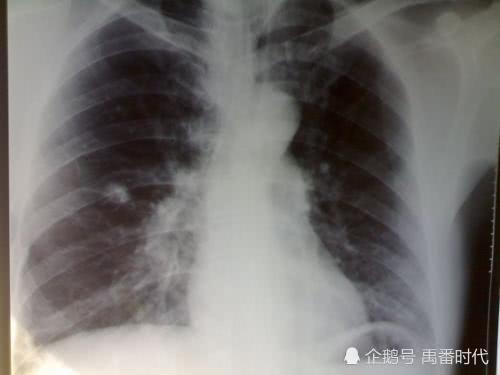
Pneumoconiosis and lung cancer actually differ in their pathogenesis, symptoms and manifestations, and in various other aspects. Pneumoconiosis, for example, occurs when dust and particulate matter are deposited in the alveoli and cause pulmonary fibrosis, a process that takes a long time to develop. Lung cancer, on the other hand, is the evolution of various lung diseases into a malignant tumor. Pneumoconiosis does not metastasize, whereas malignant tumors do metastasize, and pneumoconiosis can also develop into lung cancer, but lung cancer is decay, the final destination of the disease.
In terms of symptoms and manifestations, in the early and middle stages of pneumoconiosis, or when it is complicated by other diseases, symptoms such as cough, sputum production, chest pain, difficulty breathing and hemoptysis may appear; in severe and late stages, symptoms such as facial and neck edema, hoarseness and shortness of breath may be added. Early-stage lung cancer brings symptoms such as cough, low-grade fever, chest distension and pain, and blood-streaked sputum; the late stage mainly refers to the symptoms added after lung cancer metastasizes to other organs or systems.
Small rounded opacities
On chest X-rays and other examinations, pneumoconiosis and lung cancer also differ from each other. In typical silicotic pneumoconiosis, for example, the two lung fields initially show basically symmetrical small rounded opacities, which are more obvious on the outer side. They generally do not involve the lung apices; if an opacity appears at a lung apex, it is usually pneumoconiosis complicated by pulmonary tuberculosis. As it develops, the small rounded opacities also appear in the middle and lower lung fields, and the opacities increase in number, become larger and become denser. When the complications of pneumoconiosis reach a severe stage, densely packed opacities like a blizzard fill both lungs, and the lung markings also become interrupted or hard to distinguish.
Lung tumors, that is, lung cancer, generally fall into three types: central, peripheral and diffuse. On chest X-rays, apart from the diffuse type, which is easily confused with pneumoconiosis, the central and peripheral types are both clearly distinguishable from pneumoconiosis. In typical central lung cancer, the presentation is a mass at the hilum protruding into the lung; in the later severe stage, the mass will include metastatic lymph nodes and may also be complicated by obstructive pneumonia and atelectasis.
Lung cancer-like rounded nodule
Peripheral lung cancer on chest fluoroscopy is mostly a round-like shape, with nodules or masses in the lung, and the margins are lobulated with small spicules and no calcification; the diffuse type mostly occurs with malignant transformation of bronchioloalveolar cells, so extensive nodular or infiltrative lesions form in both lungs. The nodules are about 1 to 5 mm in size, are distributed with uniform density and clear outlines, and tend to coalesce, but are often asymmetrical, with some lesions being denser. CT examination can detect low-density malignant tumors hidden beside the heart and beside the spine.
If you have any questions or need help, you can call 01062170649 13269852331